Navigating a diagnosis of a Haemangioma can feel overwhelming, but understanding the natural progression of these vascular growths is the first step toward effective health management and peace of mind. This article provides evidence-based insights into how these conditions are diagnosed, the various treatment pathways available, and what you can realistically expect during the recovery journey. By clarifying the difference between benign lesions and vascular malformations, we aim to equip you with the practical knowledge needed to make informed decisions for yourself or your child.
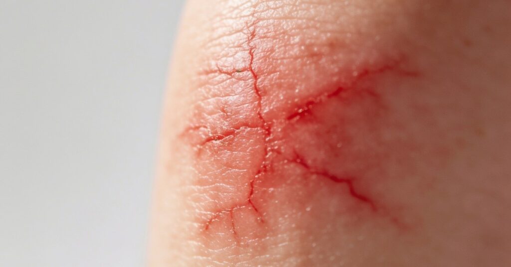
A Haemangioma is a benign tumour of endothelial cells that typically manifests shortly after birth as a red or bluish mark, often undergoing a rapid growth phase followed by a long period of natural regression. While these growths are common, they are fundamentally different from vascular malformations, which are structural defects present from birth that grow in proportion with the child’s body. Distinguishing between these two is critical for medical management, as Haemangiomas follow a specific life cycle of proliferation and involution, whereas malformations remain lifelong structural anomalies.
Understanding the Nature and Varieties of Haemangioma of Infancy and Congenital Haemangioma
Haemangiomas are classified primarily by their anatomical depth and growth characteristics, which helps a Dermatologist determine the most appropriate care strategy. Superficial Haemangiomas, often referred to as strawberry Haemangiomas, present as bright, raised red marks on the skin’s surface, while deep Haemangiomas develop beneath the dermis and frequently appear as a bluish swelling or mass.
Beyond these common presentations, there are several distinct types that require careful identification. Understanding the specific subtype is essential because it dictates the potential for complications and the necessity of medical intervention. Whether dealing with superficial lesions or more complex internal growths, categorisation is the foundation of patient care.
- Superficial: Strawberry-like, raised red marks.
- Deep: Sub-dermal bluish swellings.
- Mixed/Combined: A blend of both surface and deep tissue involvement.
- Congenital: Fully formed at birth (RICH, NICH, or partially involuting).
- Other types: Hepatic, cherry angiomas (Campbell de Morgan spots), and lobular capillary Haemangiomas.
Distinguishing Haemangiomas from Vascular Malformations
The primary difference between these two conditions is that Haemangiomas are biological tumours that exhibit a rapid growth phase between 0–6 months of age, whereas vascular malformations are static structural defects present at birth that grow proportionally with the child’s body. Because Haemangiomas of Infancy rarely appear at birth, their sudden emergence in the weeks following delivery is a hallmark clinical indicator that helps medical professionals differentiate them from congenital malformations.
| Feature | Haemangioma | Vascular Malformation |
|---|---|---|
| Presence at Birth | Rarely | Always |
| Growth Pattern | Rapid (0-6 months) | Proportional to body |
| Nature | Benign tumour | Structural defect |
Epidemiological data further helps in the diagnostic process, as venous malformations have an estimated population incidence of 1-2%. For those seeking expert evaluation, the Leeds Teaching Hospitals NHS Trust vascular clinics operate 09:00-16:00, Monday to Friday, providing a dedicated environment for the assessment of complex vascular anomalies. Knowing these differences helps parents avoid unnecessary panic when a mark appears in the early stages of life.
Clinical Diagnosis and Assessment of Infantile Signs and Symptoms
Diagnosis of an Infantile Haemangioma is primarily achieved through a comprehensive physical examination and a detailed review of the patient’s medical history. When a lesion is located in a complex area, clinicians rely on a structured approach to confirm the nature of the growth.
- Initial physical inspection of skin and eyes.
- Review of medical history to establish growth timeline.
- Imaging such as Doppler ultrasound, MRI, or abdominal ultrasound for deep lesions.
- Biopsy or GLUT-1 protein staining for diagnostic confirmation.
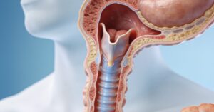
- Laryngoscopy if airway involvement is suspected.
These investigative steps are vital for ruling out other vascular conditions that might require different treatment strategies. By utilising advanced imaging like MRI or simple clinical assessments, doctors can map the extent of the tissue involvement, ensuring that no vital structures are compromised by the growing mass.
Natural Progression and Resolution Expectations for Haemangiomas
The majority of Haemangiomas will resolve on their own without the need for invasive medical intervention. Clinical evidence confirms that ninety percent (90%) of these lesions follow a self-limiting path, where they gradually shrink and fade over several years.
The timeline for this natural regression is predictable, with nearly all Haemangiomas resolving by age 10. Many parents observe significant improvements earlier, as up to 50% of these growths disappear by the time the child reaches age 5. While it is important to acknowledge that in some rare instances a Haemangioma may never disappear completely, the natural history of the condition is overwhelmingly favourable, requiring only observation in uncomplicated cases.
Medical Management and Treatment Protocols for Haemangiomas
Treatment for Haemangiomas is reserved for complicated or high-risk cases, with oral Propranolol serving as the first-line therapy for lesions that threaten vital functions or cause severe ulceration. HEMANGEOL is the specific FDA-approved oral beta-blocker used for this purpose, and the treatment duration is typically 6 to 18 months to ensure the lesion remains suppressed throughout its proliferative phase.
Important: Always keep a simple diary of your child’s medication schedule and any observed reactions; in my experience, having this documentation makes consultations with your specialist far more productive.
When the condition requires alternative or adjunctive approaches, specialists may utilise timolol gel, a topical beta-blocker effective for thin, superficial lesions. For second-line management, corticosteroids such as triamcinolone or betamethasone injections are available. In rare, life-threatening cases that do not respond to conventional therapies, interferon α and vincristine are utilised. Finally, for residual deformities or lesions that remain unresponsive to medication, surgical excision is performed to restore normal skin appearance and function. Additionally, pulsed-dye and Nd:YAG lasers are frequently employed to treat ulcerated lesions and address any residual skin discoloration left after the growth has regressed.
Frequently Asked Questions
Can a Haemangioma be inherited?
Haemangiomas are generally not considered hereditary, as they usually occur sporadically without a clear familial link. Most research suggests they are the result of localised factors during early development rather than genetic inheritance.
Is it safe to touch or clean a Haemangioma?
Yes, it is perfectly safe to touch or clean the area gently with mild soap and water during routine bathing. Avoid scrubbing or applying harsh pressure, as this may irritate the delicate surface of the lesion.
Why do some Haemangiomas develop ulcers?
Ulceration occurs when the rapid growth of the lesion causes the overlying skin to become thin and fragile, breaking down due to moisture or friction. This complication requires prompt medical attention to prevent infection and promote healing.
Are there dietary restrictions for a child with a Haemangioma?
There are no specific dietary restrictions required for a child simply because they have a Haemangioma. However, if your child is prescribed medication like Propranolol, your doctor may provide specific instructions regarding feeding schedules to ensure the medication is absorbed correctly.
Trust that your child’s growth will likely fade with time, and stay consistent with any prescribed treatment to ensure they reach that milestone safely.